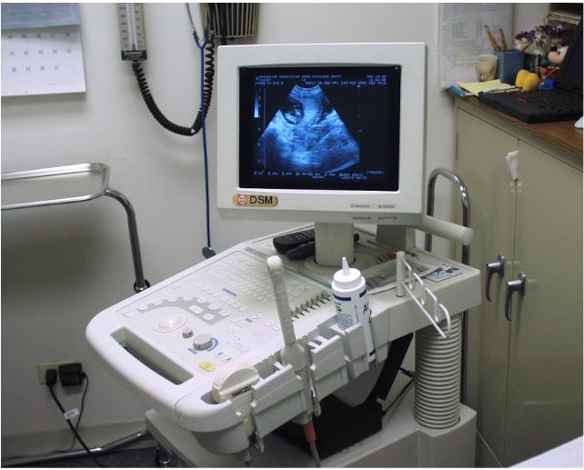
Ultrasonography uses high-frequency sound (ultrasound) waves to produce images of internal organs and other tissues. A device called a transducer converts electrical current into sound waves, which are sent into the body’s tissues. Sound waves bounce off structures in the body and are reflected back to the transducer, which converts the waves into electrical signals. A computer converts the pattern of electrical signals into an image, which is displayed on a monitor and recorded on film, on videotape, or as a digital computer image. No x-rays are used.
What are the Procedure of Ultrasonography?
If the abdomen is being examined, people may be asked to refrain from eating and drinking for several hours before the test. Usually, the examiner places thick gel on the skin over the area to be examined to ensure good sound transmission. A handheld transducer is placed on the skin and moved over the area to be evaluated. To evaluate some body parts, the examiner inserts the transducer into the body—for example, into the vagina to better image the uterus and ovaries or into the anus to image the prostate gland. To evaluate the heart, the examiner sometimes attaches the transducer to a viewing tube called an endoscope and passes it down the throat into the esophagus. This procedure is called transesophageal echocardiography.
We are Committed to
Excellence in diagnostic care
We have experienced as well as highly qualified doctor's. Therefor we have excellent Diagnose Care.
Reporting & Supports
Most of the larger laboratories have fully automated equipment and quality and statistical analyses are run daily on them.
Computerised Equipments
We believe in latest and advanced technology so that we get accurate result's. We have fully computerized equipment's.